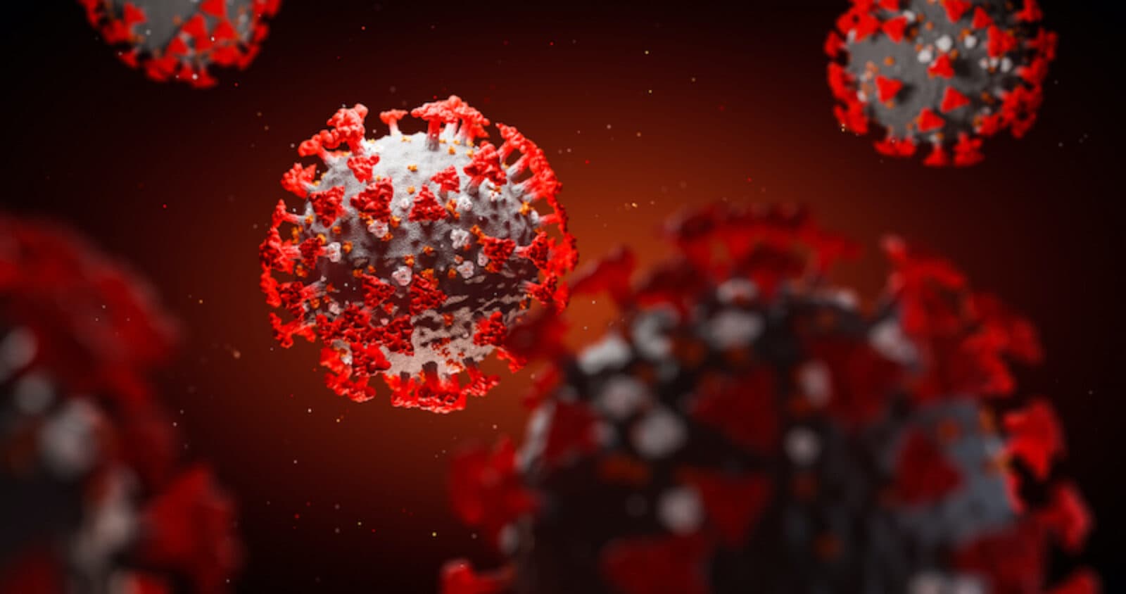
Closing in on the Coronavirus
by Alan S. Brown
Pamela Bjorkman's collaboration with Caltech's KNI could help unlock the coronavirus' smallest secrets

The Author
The Researcher
As COVID-19 swept across the United States, California Institute of Technology biologist Pamela Bjorkman pivoted her lab to study the new coronavirus. She was not looking for a quick cure. Yet the discoveries she makes may help protect us against this pandemic--and the others likely to follow in the future.
Bjorkman's work is not about quick fixes. Her team probes the fundamental mechanisms that the body's immune system uses to fight off viruses.
“What we discover helps us think about how to design therapeutics that could possibly work against viruses and vaccines for diseases that we cannot yet prevent,” she said.
One of those viruses is HIV, which causes AIDS. Bjorkman has studied it for over a decade, investigating the nanoscale siege warfare as HIV invades our bodies and our immune system throws up defenses to keep it out.
What sets Bjorkman's work apart is that she also looks at HIV in tissues rather than just cells. Most tissues contain many different types of cells, and each type interacts with HIV differently. By measuring and tracing those interactions, she develops a deeper understanding of HIV's behavior--and, perhaps, its vulnerabilities--in the real world.
This calls for specialized equipment, such as those operated by Caltech's Kavli Nanoscience Institute. Thanks to an unusual partnership--and a broken collarbone--Bjorkman and KNI have modified a KNI transmission electron microscope (TEM) to take three-dimensional photos of viruses in tissues.
Now, she is taking the methods she used to study HIV and is turning it on SARS-CoV-2.
HIV
HIV is a relentless adversary, and Bjorkman has spent the past 10 years studying it. Its key strength is its breakneck mutation rate. While influenza mutations might produce three or four major new circulating strains each year, HIV mutates so quickly that there are now estimated to be 10 quadrillion—10 million billion—variants loose in the world today.
“There are a massive number of strains in a single person infected with HIV,” she said. “Even if you could protect against 10 percent of them, it’s not going to help.”
Instead, patients must take a variety of antiviral cocktails to control the infection for the rest of their lives. If they stop, the virus will spread.
To attack HIV, Bjorkman needs a different strategy. First, she wants to identify those HIV features that provoke immune responses, such as spikes on HIV’s outer layer (smaller versions of those in the coronavirus crown). Knowing which spikes most HIV variants have in common could help craft a treatment that will kill the virus.
Second, she needs to understand how HIV actually behaves in the body. To do this, she must look at tissues that have a variety of cells. Then she can see which cells HIV targets first, where it hides, how it spreads.
It is the same strategy she plans to use to learn more about the coronavirus' vulnerability. To do this, Bjorkman turned to the Kavli Nanoscience Institute's advanced transmission electron microscope.

Looking Closely
With hundreds of times more resolution than optical microscopes, electron microscopes revolutionized biology. Yet when it comes to looking at tissues, they also have limitations that became clear to Bjorkman when she broke her collarbone several years ago.
“The doctors took a two-dimensional X-ray and told me it had healed, but I was positive it had not,” she related. “When they took a three-dimensional CAT scan, they saw the broken edges were far apart. They couldn’t tell from the X-ray because, when you look at from it from the top, the broken pieces overlapped and looked like they had mended.
“That’s an example of what can go wrong if you take a 300-nanometer slice of a tissue and put it in an electron microscope. The organelles may look like they are on top of each other, but they’re not. Or you might see a narrow channel, but you can’t tell where it goes.”
Bjorkman’s solution was to modify the KNI microscope to tilt the TEM’s stage, which holds the sample. Just like a CAT scan, the tilting stage lets the electron beam create images from a variety of different angles. An algorithm then stitches those images together into a three-dimensional reconstruction of the sample’s structure.
The collaboration with KNI cost hundreds of thousands of dollars and took nearly one year for TEM engineers to complete. Once done, it gave Bjorkman and Mark Ladinsky, a research specialist in her lab, an instrument that could image tissue.
This led to surprising discoveries in samples from infected bone marrow. It is well known, for example, that HIV targets the immune system’s T cells, which are sometimes eaten by other white blood cells called macrophages. The surprise is that once ingested, HIV can escape into the extracellular space through narrow channels that access the macrophage’s exterior. Bjorkman and Landinsky also learned which types of cells HIV attacks early and which ones come later.
Understanding how the virus behaves in its natural state may suggest new ways to fight the virus. Bjorkman hopes to make similar discoveries about the novel coronavirus as well.
In many ways, the coronavirus is an easier target, Bjorkman said. “It mutates about as much as measles virus, and there has been an effective vaccination against measles that was made by conventional methods 50 years ago,” she said.
Now, she is ready to tackle the coronavirus. Fortunately, her lab has special permission from Caltech to remain open. The TEM is classified as a clean room, and all samples are treated to prevent them from being infectious. The only question left is where to look. That is still an unknown.
“How do you know what you’re going to find until you look at the virus—where it is, what kind of cells it’s coming out of, how it affects the gut and lung,” she said. “I don’t know what we are looking for yet. Perhaps we’ll be able to look at tissues from test animals treated with different drugs, and what we see may suggest other possible treatments. In biology, you often cannot ask a particular question until you look at a structure and see what it looks like.”
Which is exactly what fundamental research should do.