Engineers Develop Most Efficient Red Light-Activated Optogenetic Switch for Mammalian Cells
(Originally published by UC San Diego)
March 5, 2018

A team of researchers has developed a light-activated switch that can turn genes on and off in mammalian cells. This is the most efficient so-called “optogenetic switch” activated by red and far-red light that has been successfully designed and tested in animal cells—and it doesn’t require the addition of sensing molecules from outside the cells.
The light-activated genetic switch could be used to turn genes on and off in gene therapies; to turn off gene expression in future cancer therapies; and to help track and understand gene function in specific locations in the human body.
The team, led by bioengineers at the University of California San Diego, recently detailed their findings online in ACS Synthetic Biology.
“Being able to control genes deep in the body in a specific location and at a specific time, without adding external elements, is a goal our community has long sought,” said Todd Coleman, a professor of bioengineering at the Jacobs School of Engineering at UC San Diego and one of the paper’s corresponding authors. “We are controlling genes with the most desirable wavelengths of light.”
The researchers’ success in building the switch relied on two insights. First, animal cells don’t have the machinery to supply electrons to make molecules that would be sensitive to red light. It’s the equivalent of having a hair dryer and a power outlet from a foreign country, but no power cord and no power outlet adapter. So researchers led by UC San Diego postdoctoral researcher Phillip Kyriakakis went about building those.
For the power cord, they used bacterial and plant ferredoxin, an iron and sulfur protein that brings about electron transfer in a number of reactions. Ferredoxin exists under a different form in animal cells, which isn’t compatible with its plant and bacteria cousin. So an enzyme called Ferredoxin-NADP reductase, or FNR, played the role of outlet adapter.
As a result, the animal cells could now transfer enough electrons from their energy supply to other enzymes that can produce the light-sensitive molecules needed for the light-activated switch.
The second insight was that the system to make light-sensitive molecules needed to be placed in the cell’s mitochondria, the cell’s energy factory. Combining these two insights, the researchers were able to build a plant system to control genes with red light inside animal cells.
Red light is a safe option to activate genetic switches because it easily passes through the human body. A simple way to demonstrate this is to put your hand over your smart phone’s flashlight while it’s on. Red light, but not the other colors, will shine through because the body doesn’t absorb it. And because it’s not absorbed, it can actually pass through tissues harmlessly and reach deep within the body to control genes.
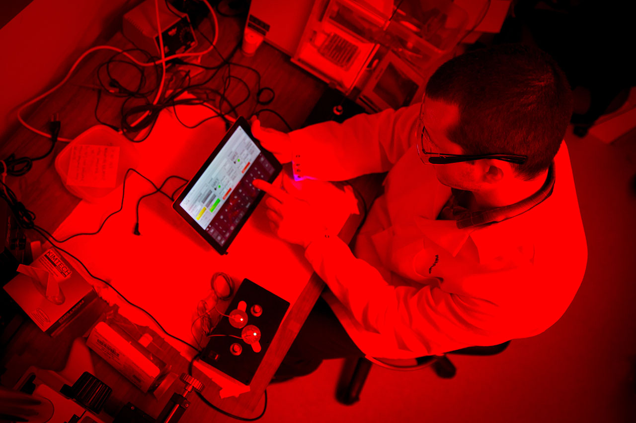
Bioengineers built and programmed a small, compact tabletop device to activate the switch with red and far-red light. The tool allows researchers to control the duration that the light shines, down to the millisecond. It also allows them to target very specific locations. Researchers showed that the genes turned on by the switch remained active for several hours in several mammalian cell lines even after a short light pulse.
The team recently received an internal campus grant to use the method to control gene activation in specific regions of the brain. This would allow them to better understand gene function in a variety of neurological disorders.
The researchers patented the use of ferredoxins and FNR to target the enzymes needed to make light-activated molecules. The technology is available for licensing.
Importantly, insights about how to produce plant molecules in animal cells could also one day enable production of other molecules that can lead to the cultivation of plants that do not need fertilizer and make biofuel production more efficient.
The study was supported by the Kavli Institute for Brain and Mind at UC San Diego and the Salk Institute, the National Science Foundation, and the National Institutes of Health.
The research team included researchers from the Division of Biological Sciences, the Neurosciences Graduate Program and the School of Medicine at UC San Diego, as well as researchers at Quinnipiac University and the University of Iowa.