Insights into the Circuits that Make Memories
by Lindsay Borthwick
Research highlights from Kavli Neuroscience Institutes

The Author
The brain’s memory system is complex, and the focus of our scientific highlights this month. A recent study identifies a long-range circuit that illuminates how the prefrontal cortex exerts top-down control over the hippocampus, the seahorse-shaped region vital to forming new memories and to navigation. The second study identifies a mechanism for age-related memory loss rooted in the CA3 region of the hippocampus. Another research team explores the development of the nearby entorhinal cortex, which together with the hippocampus modulates episodic memory that allows us to recall everyday events and experiences. And, finally, we celebrate the paradigm-shifting work of a scientist who has used mathematics to describe how brain cells can encode new memories while preserving old ones.
A long-distance relationship
Researchers at the University of California, San Francisco (UCSF) have identified a brain circuit that may shape what information we should focus on in the environment, and what we should ignore. It links the prefrontal cortex (PFC) to the hippocampus, forming a long-range connection that has not yet been described. The PFC is an area that guides thought, attention, behavior and emotion. The hippocampus is involved in memory and spatial cognition. “This circuit is a gateway to understanding how the brain allows the prefrontal cortex to exert top-down regulation of other parts of the brain,” said senior author and member of the Kavli Institute for Fundamental Neuroscience at UCSF. Vikaas Sohal, M.D., Ph.D., in a news article. Ruchi Malik, Ph.D., the study’s lead author, uses a parking lot as an example of how the newly discovered circuit might influence our behavior. “To remember where you parked, the PFC would tell the hippocampus to selectively pay attention to landmarks, and then recall and seek out those landmarks when you return,” she said. In future experiments, the team would like to investigate what happens when the circuit malfunctions, including whether it plays a role in dementia or psychiatric disorders related to attention and memory.

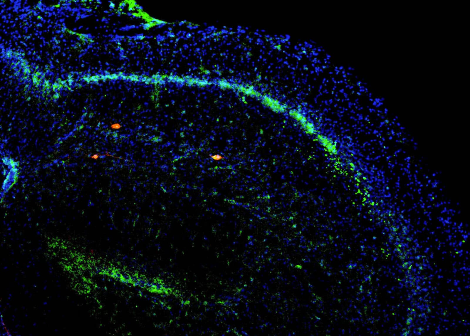
A memory-loss mechanism
The brain performs different memory functions, even in a single region. The CA3 area of the hippocampus is known for its role in pattern completion and pattern separation, two complimentary processes that help us makes sense of our daily experiences. Neuroscientist James Knierim, a member of the Kavli Neuroscience Discovery Institute at Johns Hopkins University, has shown that hyperactivity of neurons in the CA3 contributes to age-related memory loss by favoring one of these processes over the other. But the CA3 region of the brain isn’t homogenous. The cells involved in pattern completion are more common at one end of CA3, while those responsible for pattern completion are more prevalent at the other end. In a series of experiments in rats, Knierim and his team showed that with age, the neurons responsible for pattern completion become hyperactive, creating an imbalance in the brain’s memory center. This may explain the common memory lapses associated with aging. "We all make these mistakes, but they just tend to get worse with aging," Knierim said in a news article. Michaela Gallagher, another Kavli Institute member, contributed to the study.
Say it with your eyes
What happens in the brain when you make eye contact with another person? And how do we extract meaning from such a brief connection? Neuroscientists affiliated with Yale’s Kavli Institute for Neuroscience have charted how brain cells respond when two animals spontaneously make eye contact. They found widespread neuronal activity in multiple brain areas — activity that provides clues about how the brain assesses the meaning of a gaze. Cells were active in the prefrontal cortex and in the amygdala, an area of the brain that is involved in emotion. Senior author Steve Chang said the widespread activity shows just how important eye contact is. Social gaze interaction likely serves a critical role in shaping social connectedness, he said, and the PFC-amygdala networks might make that happen. Siqi Fan and Olga Dal Monte co-led the study, which was published in the journal Cell.
Memory sleuth
For most of his career, Menno Witter, a group leader at the Kavli Institute for Systems Neuroscience, has been mapping a brain region called the entorhinal cortex (EC) and studying its function. The EC nestles up against the hippocampus in the temporal lobe. Together, the two structures form the episodic memory system, which allows us to consciously remember everyday events and experiences and use them to make predictions about the future. Over the years, Witter, with his students and collaborators, has identified many of the cell types in the EC and their wiring, and showed that different parts of the EC perform different functions. In May, the Institute of Neuroscience at UC Louvain recognized Witter’s scientific achievements with an honorary degree, however, his exploration of the EC is far from complete. He recently contributed to the first detailed study of the human EC during prenatal development, with his Kavli Institute colleague Giulia Quattrocolo and first author Goran Šimić of the Croatian Institute for Brain Research. The research is an important step toward understanding how the EC develops and matures, when it begins participating in memory processes, and when it starts working with the hippocampus to encode episodic memories.
A Prize for Computational and Theoretical Neuroscience
Congratulations to Larry Abbott, Ph.D. (Columbia University), Emery Neal Brown, M.D. (Massachusetts Institute of Technology), Ph.D., Terrence Sejnowski, Ph.D. (Salk Institute for Biological Studies), and Haim Sompolinsky, Ph.D. (Hebrew University of Jerusalem), who were awarded the 2022 Gruber Neuroscience Prize “for their pioneering contributions to computational and theoretical neuroscience.” By applying their knowledge of mathematics, statistics, physics and machine learning, the prizewinners have generated new theories, models and tools that have transformed experimental neuroscience and unlocked new insights into the brain function. Abbott and Sejnowski have both been instrumental to the development of the Kavli Neuroscience Institutes: Abbott is co-director of the Kavli Institute for Brain Science
at Columbia; and, Terrence Sejnowski is a longtime member of the Kavli Institute for Brain and Mind at the the Salk Institute for Biological Studies. The prize recognizes Abbott’s research on modelling neurons and neural networks, including his landmark studies on how neurons adjust the strength of their connections in support of learning and memory. Sejnowski was a pioneer in the field of artificial intelligence and has made numerous contributions to it, including the development of the first unsupervised learning algorithm for independent component analysis, an analysis technique used in brain imaging.
Larry is willing to deal with the messiness of real neuroscience data, and work with those limitations. Theory is beautiful and internally consistent. Biology, not so much.
Cori Bargmann in a 2014 New York Times’ article about Larry Abbott’s research