Listening to the Brain
by Lindsay Borthwick
Partnerships between engineers, doctors and neuroscientists open up new ways to listen to the brain

The Author
A trio of studies from researchers at the Kavli Neuroscience Institutes highlight partnerships between engineers, doctors and neuroscientists intent on listening to the brain. They are working to develop sensors that can record brain activity at high-resolution—for research and treatment purposes. Read on to learn how brain data is being used to fine tune deep brain stimulation for Parkinson’s disease, create better brain maps and work out how the hippocampus functions.
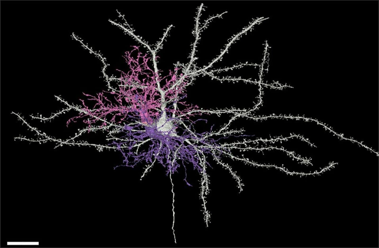
Cellular Fireworks
This stunning 3D reconstruction, seen above, of a brain cell is courtesy of the Allen Institute for Brain Science and a team of collaborators, including Dwight Bergles, director of the Kavli Neuroscience Discovery Institute at Johns Hopkins University. The cell? It is an oligodendrocyte precursor cell, or OPC, in the mouse visual cortex, painstakingly piece together at the ultrastructural level from electron micrographs. OPCs eventually give rise to oligodendrocytes, the large glial cells that produce myelin, which ensheaths and insulates neuronal axons. In a new preprint posted to bioRxiv, the team of collaborators report the discovery that oligodendrocytes, or OPCs, help refine neural circuits during the later phases of cortical development, alongside better-known circuit remodeling cells like microglia and astrocytes. In fact, the team found that OPCs contain greater numbers of phagolysosomes, cellular compartments that digest debris and portions of neurons, than microglia, “raising the possibility that they are responsible for a great amount of [synaptic] pruning at this stage of maturation of the cortex,” write the authors.
Neurostimulation Toolkit
A new Nature Biotechnology paper highlights what can be achieved when cutting-edge neurotechnology hits the clinic. For the first time, a team led by Philip Starr at the University of California, San Francisco (UCSF) continuously recorded and analyzed brain activity over extended periods of time from people with Parkinson’s disease as they went about their daily lives. Starr’s team also collected data about their movements using a wearable wristband. The brain activity and movement data are streamed in real time and can be used to fine tune neurostimulation and achieve a level of personalized treatment that hasn’t been possible for deep-brain stimulations before.
Starr is also leading the Open Mind Consortium, a multidisciplinary partnership to develop open-source technology platforms for implantable brain stimulation devices, which is funded by the US BRAIN Initiative as part of its goal of moving new neurotechnologies from the lab and into the clinic. In a news article about the consortium, John Ngai, Director of the US BRAIN Initiative said, “Dr John Ngai, director of the NIH BRAIN Initiative, said: ‘As we move toward modulating brain circuitry in real-time, the increasing complexity of the technology requires collaboration among neuroscientists, computer scientists, and engineers to develop safe and effective interventions that also adhere to neuroethical principles.” Starr is a member of the Kavli Institute for Fundamental Neuroscience at UCSF.
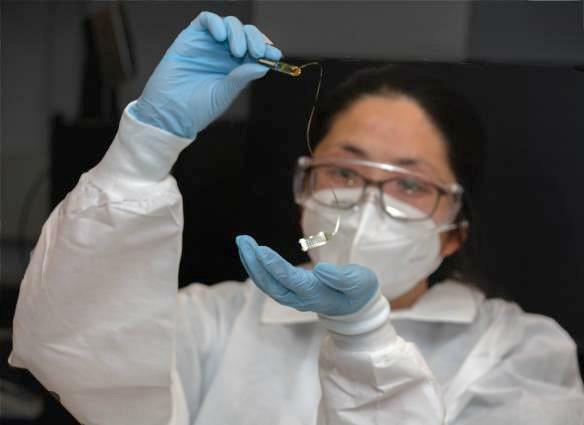
Better Brain Maps
New sensors created at the University of California, San Diego (UC San Diego) sit on brain’s surface and allow surgeons to create higher-resolution brain maps.
Neurosurgeon Wilder Penfield famously developed the “Montreal procedure" for the treatment of epilepsy, in which he stimulated parts of the cortex during surgery, searching for and then removing the scar tissue causing patients’ seizures. More than 50 years later, researchers are developing new tools to create brain maps specific to each person which can guide surgical teams as they treat epilepsy or remove brain tumors. A team from the University of California, San Diego, recently crafted flexible sensors that sit directly on the brain’s surface and record brain activity that can be used to produce high-resolution brain maps. The research is a collaboration between clinicians and engineers, namely Eric Halgren, a professor of neurosciences and radiology at UC San Diego Health, and electrical engineering professor Shadi Dayeh. The new devices have a density of sensors and are made for a more supple material that conforms to the surface of the brain. Halgren is a member of the advisory board of the Kavli Institute for Brain and Mind at UC San Diego.
Wave Reading
In yet another feat of engineering for neuroscience, researchers at Lawrence Livermore National Laboratory (LLNL) and the University of California, San Francisco (UCSF) have developed thin-film electrodes and used them to record traveling waves in the human brain. The new device, which is smaller than a dime, enabled neuroscientists to record from the surface of the hippocampus, a brain region involved in learning and memory, in patients undergoing epilepsy surgery. The results were intriguing: The researchers confirmed the existence of traveling waves and found that the waves move up and down the hippocampus.
“This ‘two-way street’... is a big deal because we believe this may be a fundamental mechanism of how the hippocampus acts as a major hub of information and memory processing for many other brain regions. In other words, the direction the wave is moving across the hippocampus may be a biomarker reflecting distinct neural processes as different circuits engage and disengage,” said the paper’s lead author, UCSF neurologist Jon Kleen.
The senior authors are Razi Haque, LLNL’s Implantable Microsystems Group Leader Razi Haque and UCSF neurosurgeon Edward Chang. They are both members of the Kavli Institute for Fundamental Neuroscience at UCSF.
Close up
A new study is probing the characteristics of live human pyramidal cells, the major class of neurons in the neocortex, to try to understand what makes human brain cells, well, “human.” A team of researchers based at the University Health Network and the Centre for Mental Health and Addiction in Toronto, studied more than 200 neurons from 61 people who were undergoing epilepsy surgery — ”one of the largest studies of the electrophysiological diversity of human neocortical pyramidal cells to date.” While the researchers don’t hail from a Kavli Institute, they were partly supported by a grant from The Kavli Foundation through Neurodata Without Borders (NWB), The resulting dataset, which uses the NWB:Neurophysiology data format, is one of the largest datasets of its kind. The research revealed, among other insights, massive diversity among pyramidal neurons as well as major differences between pyramidal cells in humans and other mammals.