Take a Tour of the Brain
by Lindsay Borthwick
Highlights from Kavli Neuroscience Institutes

The Author
From the brainstem to the cerebral cortex, researchers at the Kavli Neuroscience Institutes are probing the brain in new ways. We begin with the work of Kevin Yackle at the University of California, San Francisco, who studies the neuroscience behind breathing. It covers a couple of fascinating discoveries by Attila Loconszy of the Kavli Institute for Brain Science at Columbia University, whose research on the hippocampus demonstrates how much we still have to learn about memory. Moving from the deep brain out toward the cerebral cortex, we visit Shadi Dayeh at the Kavli Institute for Brain and Mind at the University of California, San Diego. He builds sensors that sit on the brain's surface and capture cortical activity in incredible detail, guiding brain surgeons and neuroscientists striving to understand cognition. Enjoy the journey.
The rhythm of our breath
We breathe automatically, about 12 times per minute. We also speak and talk without much thought, rhythmically producing a string of syllables between each breath. According to new research, led by Kevin Yackle, MD, Ph.D., at the University of California, San Francisco (UCSF), that rhythm may be driven by cells in the brainstem, a region that regulates many life-sustaining functions. Yackle's team studies breathing: How we do it; how it changes during disease; and, how it is coordinating with other behaviors such as swallowing, coughing and talking. They recently reported the discovery of clusters of cells in the brainstem of mice that appear to coordinate the rhythms of breathing and vocalizations. The finding is exciting because it may help settle a debate about whether speech is learned or innate. Yackle's discovery suggests nature plays a significant part: "This suggests that there is a hardwired network of neurons that is fundamental to speech," he told a reporter from NPR. Yackle suspects similar cells exist in humans and could explain why babies can cry from the moment they are born and why many human languages have a characteristic rhythm. Yackle is a member of the Kavli Institute for Fundamental Neuroscience at UCSF.
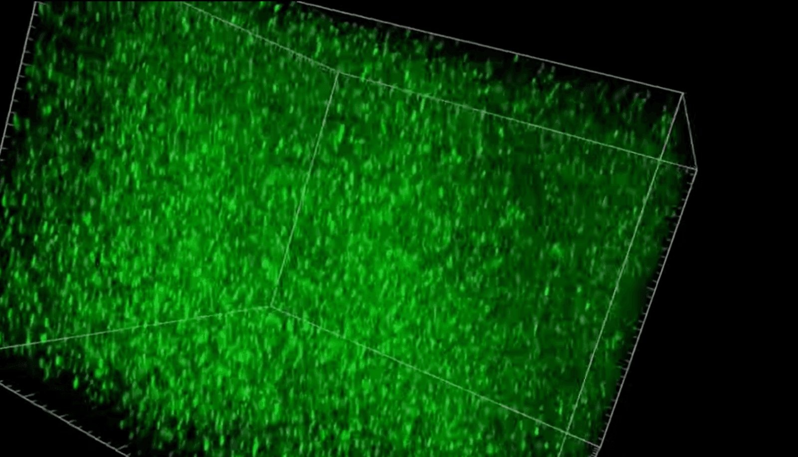
Seeing synapses in a new way
The brain's ability to learn and lay down new memories hinges on the crosstalk between neurons. Those conversations occur at synapses, the physical connections between neurons where information is processed and stored. Over several decades, researchers have zoomed in on synapses and worked out many of the details of synaptic function. But, until now, they haven't been able to pull back and see synapses in action across large areas of the brain. A new research tool, developed in the laboratory of Richard Huganir, Ph.D., former director of the Kavli Neuroscience Discovery Institute, can reveal constellations of millions of synapses in the cortex of healthy, living mice. The researchers engineered the mice to synthesize a synaptic protein receptor with a fluorescent tag. When the receptors are activated by neuronal activity, the synapses glow, allowing the researchers to watch what happens as the mice interact with the world around them, learn and lay down memories. In the future, the tool could allow researchers to study how synaptic activity changes on a massive scale during development and ageing, and for brain disorders such as Alzheimer's.
Mapping the cortex in high resolution
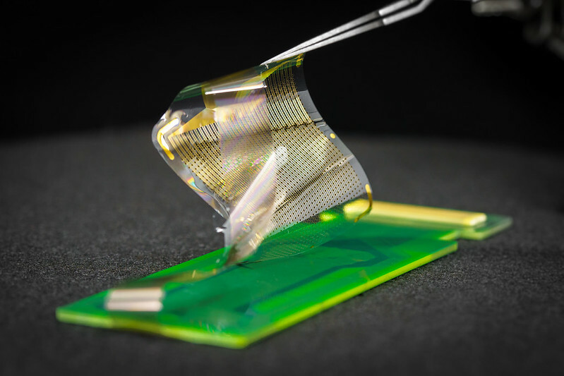
Sensors can help us measure the invisible. Shadi Dayeh, Ph.D., an electrical engineering professor at the University of California, San Diego (UCSD), builds sensors that can be applied to the brain's surface. A typical neurosurgical array contains 16 and 64 sensors. Dayeh's thin, pliable grids have 1,024 or 2,048 sensors packed into a similar area. As a result, they reveal the activity of the cerebral cortex in remarkable detail. He described these new sensors in the January issue of the journal Science Translational Medicine. Dayeh and his collaborators showed that the sensors can help surgeons operate with greater precision to remove brain tumors or misfiring tissue that causes epileptic seizures. The sensors can also yield fundamental insights into the brain. For example, for the first time, the researchers mapped a cortical column, the basic functional unit of the cerebral cortex, from the brain's surface. Dayeh is an advisory board member of the Kavli Institute for Brain and Mind at UCSD. He is leading a research team that recently received more than $12 million from the U.S. BRAIN Initiative to further develop the technology in the lab and the clinic.
The fly guy
Gaby Maimon, Ph.D., knows that insects can teach us a lot about the brain, including how it forms memories and uses those memories to guide behavior. He studies how the tiny fruit fly navigates, even in the dark, to understand the complex operations that help animals find their way in the world. In a new study, Maimon and his team describe how fruit flies calculate their direction in space. His team previously discovered a kind of internal compass, which signals the direction a fly is facing. The new research goes a step further: It describes how the insects calculate the direction they are moving, even when facing a different way. That calculation is performed by newly identified neurons that track body movement and orientation. "This is the first set of cells known to indicate which way an animal is moving in a world-centered reference frame," Maimon said in an article about the research. In collaboration with theoretical neuroscientist Larry Abbott, Ph.D., Maimon's team also showed that the flies perform a mathematical operation to figure out which way they are going from moment to moment. The calculation may serve as a blueprint for how animals with larger brains navigate. Maimon is a steering committee member of the Kavli Neural Systems Institute at The Rockefeller University, and Abbott is co-director of the Kavli Institute for Brain Science at Columbia University.
How memories are made
Attila Locoszy, MD, Ph.D., a member of the Kavli Institute for Brain Science at Columbia University, studies how the cells in the hippocampus learn and encode information. Two studies from his laboratory, recently published in Nature, demonstrate the power of new technologies to yield insights into learning and memory. In the first study, his team developed a new technique to study small groups of cells in the hippocampus. Their goal was to understand how place cells, which encode memories of locations, talk to surrounding cells as memories are formed. What they found was a surprise: Nearby excitatory cells talked to each other when neighboring place cells were active. Previously, it was thought that excitatory cells only spoke to nearby inhibitory ones. According to the authors, communicating between excitatory cells allows single place cells to reshape local circuits. The findings add to our fundamental understanding about how the brain records memories of places.
In a second study led by Loconszy, a research team looked at a different region of the hippocampus involved in learning and remembering the relationship between unrelated details, such as the name of someone we have just met. The team developed a new method to image brain cells deep within that region. They discovered the cells help the brain filter out unimportant details, contributing to its ability to learn what information matters in a new situation. The filtering process happens as memories are stabilized.